Contact us

Selected Publications

  1. The Effect of High Pressure on Polymorphs of a Derivative of Blatter’s Radical: Identification of the Structural Signatures of Subtle Phase Transitions., Crystal Growth & Design, 2023, 23(3), pp.1915-1924.

    Edward T. Broadhurst, Cameron J. G. Wilson, Georgia A. Zissimou, Mayra A. Padrón Gómez, Daniel M. L. Vasconcelos, Christos P. Constantinides, Panayiotis A. Koutentis, Alejandro P. Ayala, and Simon Parsons.

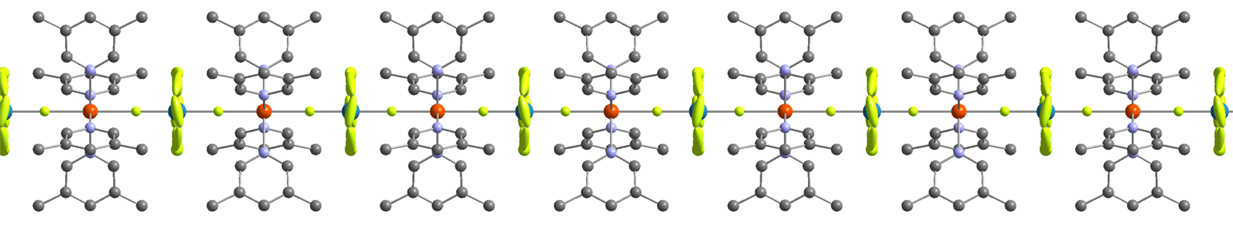
    The effect of pressure on the α and β polymorphs of a derivative of Blatter’s radical, 3-phenyl-1-(pyrid-2-yl)-1,4-dihydrobenzo[e][1,2,4]triazin-4-yl, has been investigated using singlecrystal X-ray diffraction to maximum pressures of 5.76 and 7.42 GPa, respectively. The most compressible crystallographic direction in both structures lies parallel to π-stacking interactions, which semiempirical Pixel calculations indicate are also the strongest interactions present. The mechanism of compression in perpendicular directions is determined by void distributions. Discontinuities in the vibrational frequencies observed in Raman spectra measured between ambient pressure and ∼5.5 GPa show that both polymorphs undergo phase transitions, the α phase at 0.8 GPa and the β phase at 2.1 GPa. The structural signatures of the transitions, which signal the onset of compression of initially more rigid intermolecular contacts, were identified from the trends in the occupied and unoccupied volumes of the unit cell with pressure and in the case of the β phase by deviations from an ideal model of compression defined by Birch−Murnaghan equations of state.

    DOI: 10.1021/acs.cgd.2c01422


  2. Discerning subtle high-pressure phase transitions in glyphosate. CrystEngComm, 2023, 25(6), pp.988-997.

    Cameron J. G. Wilson, Peter A. Wood and Simon Parsons

    The common garden herbicide glyphosate, N-(phosphonomethyl)glycine, has been studied between ambient pressure and 5.17 GPa using single crystal X-ray diffraction. Glyphosate forms a structure composed of layers parallel to the (1 0 −2) planes. Hydrogen bonds form along the stacking direction, which are very incompressible so that the effects of pressure are accommodated mostly within the layers. This study has confirmed two high pressure phase transitions previously observed by Raman spectroscopy, enabling the structural signatures of the transitions to be identified. Both transitions are very subtle and second order, involving changes to the way the structure responds to pressure rather than changes to the structure. The first transition occurs between 0.93–1.21 GPa and corresponds to the onset of greater compressibility within the layers. The second transition between 3.78–4.23 GPa is an intramolecular feature signalling a deformation of the molecular backbone. In the absence of a first order phase transition, the packing remains in a compressed form of its ambient pressure form up until the highest pressure measured. A reconstructive phase transition occurs at 5.98 GPa forming a polycrystalline high-pressure phase.

    DOI: 10.1039/d2ce01616h


  3. Behavior of Occupied and Void Space in Molecular Crystal Structures at High Pressure Crystal growth & design, 202222(4), pp.2328-2341.

    Cameron J. G. Wilson, Tomas Cervenka, Peter A. Wood, and Simon Parsons

    We report a Monte Carlo algorithm for calculation of occupied (“network”) and unoccupied (“void”) space in crystal structures. The variation of the volumes of the voids and the network of intermolecular contacts with pressure sensitively reveals discontinuities associated with first- and second-order phase transitions, providing insights into the effect of compression (and, in principle, other external stimuli) at a level between those observed in individual contact distances and the overall unit cell dimensions. The method is shown to be especially useful for the correlation of high-pressure crystallographic and spectroscopic data, illustrated for naphthalene, where a phase transition previously detected by vibrational spectroscopy, and debated in the literature for over 80 years, has been revealed unambiguously in crystallographic data for the first time. Premonitory behavior before a phase transition and crystal collapse at the end of a compression series has also been detected. The network and void volumes for 129 high-pressure studies taken from the Cambridge Structural Database (CSD) were fitted to equation of state to show that networks typically have bulk moduli between 40 and 150 GPa, while those of voids fall into a much smaller range, 2–5 GPa. These figures are shown to reproduce the narrow range of overall bulk moduli of molecular solids (ca. 5–20 GPa). The program, called CellVol, has been written in Python using the CSD Python API and can be run through the command line or through the Cambridge Crystallographic Data Centre’s Mercury interface.

    DOI: 10.1021/acs.cgd.1c01427


  4. A first-order phase transition in Blatter's radical at high pressure Acta Crystallographica Section B: Structural Science,2022, Crystal Engineering and Materials78(2)

    Edward T. Broadhurst, Cameron J. G. Wilson, Georgia A. Zissimou, Fabio Nudelman, Christos P. Constantinides,  Panayiotis A. Koutentis and Simon Parsons

    The crystal structure of Blatter's radical (1,3-di­phenyl-1,4-di­hydro­benzo[e][1,2,4]triazin-4-yl) has been investigated between ambient pressure and 6.07 GPa. The sample remains in a compressed form of the ambient-pressure phase up to 5.34 GPa, the largest direction of strain being parallel to the direction of π-stacking interactions. The bulk modulus is 7.4 (6) GPa, with a pressure derivative equal to 9.33 (11). As pressure increases, the phenyl groups attached to the N1 and C3 positions of the triazinyl moieties of neighbouring pairs of molecules approach each other, causing the former to begin to rotate between 3.42 to 5.34 GPa. The onset of this phenyl rotation may be interpreted as a second-order phase transition which introduces a new mode for accommodating pressure. It is premonitory to a first-order isosymmetric phase transition which occurs on increasing pressure from 5.34 to 5.54 GPa. Although the phase transition is driven by volume minimization, rather than relief of unfavourable contacts, it is accompanied by a sharp jump in the orientation of the rotation angle of the phenyl group. DFT calculations suggest that the adoption of a more planar conformation by the triazinyl moiety at the phase transition can be attributed to relief of intramolecular H...H contacts at the transition. Although no dimerization of the radicals occurs, the π-stacking interactions are compressed by 0.341 (3) Å between ambient pressure and 6.07 GPa.

    DOI: 10.1107/S2052520622000191


  5. Use of a miniature diamond-anvil cell in a joint X-ray and neutron high-pressure study on copper sulfate pentahydrate. IUCrJ. 2021;9(1).

    Giulia Novelli, Konstantin V. Kamenev, Helen E. Maynard-Casely, Simon Parsons, Garry J. McIntyre.

    Single-crystal X-ray and neutron diffraction data are usually collected using separate samples. This is a disadvantage when the sample is studied at high pressure because it is very difficult to achieve exactly the same pressure in two separate experiments, especially if the neutron data are collected using Laue methods where precise absolute values of the unit-cell dimensions cannot be measured to check how close the pressures are. In this study, diffraction data have been collected under the same conditions on the same sample of copper(II) sulfate pentahydrate, using a conventional laboratory diffractometer and source for the X-ray measurements and the Koala single-crystal Laue diffractometer at the ANSTO facility for the neutron measurements. The sample, of dimensions 0.40 × 0.22 × 0.20 mm3 and held at a pressure of 0.71 GPa, was contained in a miniature Merrill–Bassett diamond-anvil cell. The highly penetrating diffracted neutron beams passing through the metal body of the miniature cell as well as through the diamonds yielded data suitable for structure refinement, and compensated for the low completeness of the X-ray measurements, which was only 24% on account of the triclinic symmetry of the sample and the shading of reciprocal space by the cell. The two data-sets were combined in a single `XN' structure refinement in which all atoms, including H atoms, were refined with anisotropic displacement parameters. The precision of the structural parameters was improved by a factor of up to 50% in the XN refinement compared with refinements using the X-ray or neutron data separately.

    DOI: 10.1107/S2052252521010708


  6. Revealing the early stages of carbamazepine crystallization by cryoTEM and 3D electron diffraction  IUCrJ. 2021;8(6)

    Edward T Broadhurst, Hongyi Xu, Simon Parsons, Fabio Nudelman

    Time-resolved carbamazepine crystallization from wet ethanol has been monitored using a combination of cryoTEM and 3D electron diffraction. Carbamazepine is shown to crystallize exclusively as a dihydrate after 180 s. When the timescale was reduced to 30 s, three further polymorphs could be identified. At 20 s, the development of early stage carbamazepine dihydrate was observed through phase separation. This work reveals two possible crystallization pathways present in this active pharmaceutical ingredient.

    DOI: 10.1107/S2052252521010101


  7. Accurate H‐atom parameters for the two polymorphs of l‐histidine at 5, 105 and 295 K. Acta Crystallographica Section B: Structural Science, Crystal Engineering and Materials. 2021;77(5):785-800.

    Giulia Novelli, Charles J McMonagle, Florian Kleemiss, Michael Probert, Horst Puschmann, Simon Grabowsky, Helen E Maynard-Casely, Garry J McIntyre, Simon Parsons

    The crystal structure of the monoclinic polymorph of the primary amino acid L-histi­dine has been determined for the first time by single-crystal neutron diffraction, while that of the orthorhombic polymorph has been reinvestigated with an untwinned crystal, improving the experimental precision and accuracy. For each polymorph, neutron diffraction data were collected at 5, 105 and 295 K. Single-crystal X-ray diffraction experiments were also performed at the same temperatures. The two polymorphs, whose crystal packing is interpreted by intermolecular interaction energies calculated using the Pixel method, show differences in the energy and geometry of the hydrogen bond formed along the c direction. Taking advantage of the X-ray diffraction data collected at 5 K, the precision and accuracy of the new Hirshfeld atom refinement method im­ple­mented in NoSpherA2 were probed choosing various settings of the functionals and basis sets, together with the use of explicit clusters of molecules and enhanced rigid-body restraints for H atoms. Equivalent atomic coordinates and aniso­tropic displacement parameters were com­pared and found to agree well with those obtained from the corresponding neutron structural models.

    DOI: 10.1107/S205252062100740X


  8. Mapping the cooperativity pathways in spin crossover complexes. Chem. Sci., 202012, 1007-1015

    Matthew G Reeves, Elodie Tailleur, Peter A Wood, Mathieu Marchivie, Guillaume Chastanet, Philippe Guionneau, Simon Parsons

    Crystal packing energy calculations are applied to the [Fe(PM-L)2(NCS)2] family of spin crossover (SCO) complexes (PM-L = 4-substituted derivatives of the N-(2-pyridylmethylene)-4-aminobiphenyl ligand) with the aim of relating quantitatively the cooperativity of observed SCO transitions to intermolecular interactions in the crystal structures. This approach reveals a linear variation of the transition abruptness with the sum of the magnitudes of the interaction energy changes within the first molecular coordination sphere in the crystal structure. Abrupt transitions are associated with the presence of significant stabilising and destabilising changes in intermolecular interaction energies. While the numerical trend established for the PM-L family does not directly extend to other classes of SCO complex in which the intermolecular interactions may be very different, a plot of transition abruptness against the range of interaction energy changes normalised by the largest change shows a clustering of complexes with similar transition abruptness. The changes in intermolecular interactions are conveniently visualised using energy difference frameworks, which illustrate the cooperativity pathways of an SCO transition.

    DIO: 10.1039/D0SC05819J


  9. Effect of High Pressure on the Crystal Structures of Polymorphs of l-Histidine. Cryst. Growth Des. 2020, 20, 12, 7788–7804

    Giulia Novelli, Helen E Maynard-Casely, Garry J McIntyre, Mark R Warren, Simon Parsons

    The effect of pressure on the crystal structures of the two ambient-pressure polymorphs of the amino acid l-histidine has been investigated. Single-crystal diffraction measurements, up to 6.60 GPa for the orthorhombic form I (P212121) and 6.85 GPa for the monoclinic form II (P21), show their crystal structures undergo isosymmetric single-crystal-to-single-crystal first-order phase transitions at 4.5 and 3.1 GPa to forms I′ and II′, respectively. Although the similarity in crystal packing and intermolecular interaction energies between the polymorphs is remarkable at ambient conditions, the manner in which each polymorph responds to pressure is different. Form II is found to be more compressible than form I, with bulk moduli of 11.6(6) GPa and 14.0(5) GPa, respectively. The order of compressibility follows the densities of the polymorphs at ambient conditions (1.450 and 1.439 g cm–3 for phases I and II, respectively). The difference is also related to the space-group symmetry, the softer monoclinic form having more degrees of freedom available to accommodate the change in pressure. In the orthorhombic form, the imidazole-based hydrogen atom involved in the H-bond along the c-direction swaps the acceptor oxygen atom at the transition to phase I′; the same swap occurs just after the phase transition in the monoclinic form and is also preceded by a bifurcation. Concurrently, the H-bond and the long-range electrostatic interaction along the b-direction form a three-centered H-bond at the I to I′ transition, while they swap their character during the II to II′ transition. The structural data were interpreted using periodic-density-functional theory, symmetry-adapted perturbation theory, and semiempirical Pixel calculations, which indicate that the transition is driven by minimization of volume, the intermolecular interactions generally being destabilized by the phase transitions. Nevertheless, volume calculations are used to show that networks of intermolecular contacts in both phases are very much less compressible than the interstitial void spaces, having bulk moduli similar to moderately hard metals. The volumes of the networks actually expand over the course of both phase transitions, with the overall unit-cell-volume decrease occurring through larger compression of interstitial void space.

    DOI: 10.1021/acs.cgd.0c01085


  10. MrPIXEL: automated execution of Pixel calculations via the Mercury interface. J. Appl. Cryst. 202053, 1154-1162

    Matthew G Reeves, Peter A Wood, Simon Parsons

    The interpretation of crystal structures in terms of intermolecular interaction energies enables phase stability and polymorphism to be rationalized in terms of quantitative thermodynamic models, while also providing insight into the origin of physical and chemical properties including solubility, compressibility and host–guest formation. The Pixel method is a semi-empirical procedure for the calculation of intermolecular interactions and lattice energies based only on crystal structure information. Molecules are represented as blocks of undistorted ab initio molecular electron and nuclear densities subdivided into small volume elements called pixels. Electrostatic, polarization, dispersion and Pauli repulsion terms are calculated between pairs of pixels and nuclei in different molecules, with the accumulated sum equating to the intermolecular interaction energy, which is broken down into physically meaningful component terms. The MrPIXEL procedure enables Pixel calculations to be carried out with minimal user intervention from the graphical interface of Mercury, which is part of the software distributed with the Cambridge Structural Database (CSD). Following initial setup of a crystallographic model, one module assigns atom types and writes necessary input files. A second module then submits the required electron-density calculation either locally or to a remote server, downloads the results, and submits the Pixel calculation itself. Full lattice energy calculations can be performed for structures with up to two molecules in the crystallographic asymmetric unit. For more complex cases, only molecule–molecule energies are calculated. The program makes use of the CSD Python API, which is also distributed with the CSD.

    DOI: 10.1107/S1600576720008444